From the Kabat Method1 all’ R.M.P. Kabat Concept1a
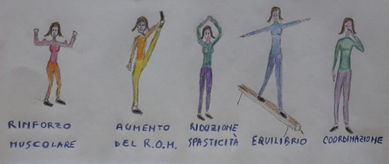
Indications: Kabat was born and developed in the field of neurorehabilitation, but his techniques are also highly indicated for orthopedic rehabilitation. The use of this method is optimal for strengthening and muscle stretching, for the increase in the width of the joint range, for the reduction of stiffness and spasticity, for coordination and balance.
The Kabat method was born in California in 1948 in the Kaiser Rehabilitation Center in Vallejo from the collaboration between the neurologist Herman Kabat and the physiotherapist Margaret Knott, to treat patients with neurological diseases. Currently this method is widely used in Europe and the two Americas. The French call it the Kabat method, the Anglo-Saxons PNF or proprioceptive neuromuscular facilitation. In Italy a variant is used which is called RMP Kabat Concept (RMP with Neurokinetic Facilitations-Kabat Concept by Giuseppe Monari where RMP stands for Progressive Modular Rebalancing), which is the one I used.
Dr.. Kabat believed it essential that the scientific knowledge acquired through basic research be used in clinical practice. His method was the fruit of this thinking, therefore it was born on the basis of the integration of neurophysiological knowledge with clinical practice, of the neurological medical approach with the rehabilitative physiotherapy approach. These partnerships have led to the development of different techniques, which together constitute the method.
The use of these techniques is aimed at muscle strengthening, to the increase in the joint range, to the reduction of stiffness and spasticity, to the improvement of coordination and balance.
Noel-Ducret suggests as best concise definition of this method a sentence extrapolated from an article by Viel E.2 : “Use of information of superficial origin (tactile) and of profound origin (joint position, stretching of tendons and muscles) for the excitation of the nervous system, which in turn causes the…musculature ". Now let's go into more specifics to get a more concrete idea of the method.
The physiotherapist administers precise stimuli to the patient, controlling intensity, duration, frequency and location according to the purpose, to facilitate the implementation of a specific motor act. These stimuli, called in jargon FACILITIES, they provide sensory information that helps the central nervous system plan and perform better movement. For the CNS, the fundamental information for this purpose is what is called proprioception . It is the ability to perceive the position of one's body in space even without the support of sight.
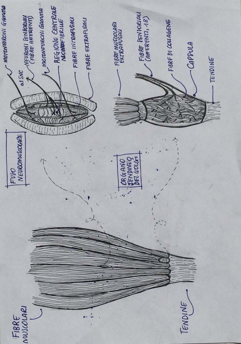
The receptors that contribute to proprioception and that are called into question by the facilitation stimuli administered by the physiotherapist are:
- I muscle receptors: Golgi tendon organs that are found in the muscle-tendon joints and that are sensitive to stretching of the tendons, both due to passive mobilization and muscle contraction. The neuromuscular spindles, sensitive to both static and dynamic muscle stretching.
· I joint receptors, present in joint capsules or ligaments, that communicate the degree of angulation in which the different body districts are found at a given moment.
· I skin receptors, sensitive to touch and any deformation of the skin.
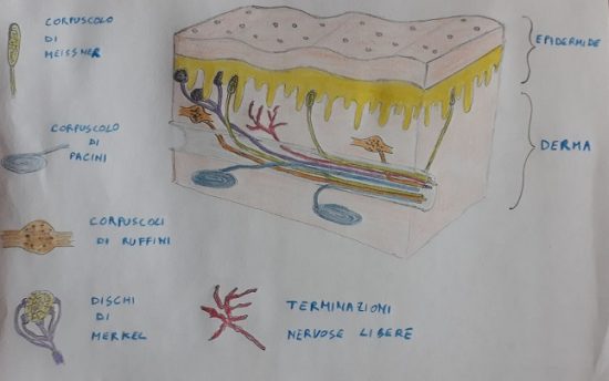
These receptors are stimulated:
· Directly from hand contact of the physiotherapist on the patient's skin
· By means of the coaptation (which means stimulating a joint in compression), extremely useful for inducing limb stability.
· With the application of a resistance to the movement (the position of the therapist's hands is crucial, as it constitutes a real guide to the direction of movement, which will take place in the opposite direction to that of the applied resistance). Resistance stimulates the recruitment of more motor units into the muscle.
· With the administration of the stimulus of stretching, which causes a brief reflex muscle contraction. The physiotherapist simultaneously asks the patient to perform a voluntary contraction in the opposite direction to that of the stretching stimulus. This contraction will benefit from the initial reflex contraction (caused by the excitation of the neuromuscular spindle due to stretching) which works as a facilitation.
· With auditory / cognitive-verbal stimulations (“verbal command”). This is a concise indication of how the patient must perform the exercise. The therapist first explains the exercise exhaustively to the patient. During the course, he then uses specific verbal commands, which indicate and stimulate a specific mode of execution: “HERE!”, "STRIP!”, "PUSH!”.
· With visual stimulations (the patient must follow the movement that the limb performs in space with his eyes).
The different techniques proposed by the Kabat method provide specific intervention methods to be used from time to time depending on the problems that need to be addressed.: improve coordination, balance, stability, resistance, muscle strength, the range of motion, reduce hypertonicity.
The neurophysiological principles on which these techniques are based are:
· The "law ofsubsequent induction”By Sherrington. The contraction of a muscle is greatest when preceded by a strong contraction of its antagonist, if the two contractions follow each other without a time interval. We can thus improve, for example, a deficient flexor movement of a limb using the force of the opposite movement, or the extensory one.
· L’mutual innervation. There are inhibitory circuits at the medullary level that cause, when a muscle (agonist) if it contracts, his antagonist releases3. We can therefore obtain the relaxation of a muscle by contracting its antagonist. The stronger the contraction of the antagonist will be, the greater the relaxation ! The inhibition mechanisms allow for a relaxation of spasticity and make it easier for the subject to perform the act.
· Irradiation: "Strong muscles are used as starters to reinforce the action of weak muscles. (…) a muscle that encounters strong resistance radiates its less vigorous brothers.4”
· Prolonged stretching. “Stretching a spastic muscle continuously (but without brutality), a progressive decrease or elimination of spasticity is achieved. The organs of the Golgi, stimulated by continued traction, produce inhibitory signals that, accumulating, They "depress" muscle tension. Just applied the traction, the motor spindles (fast running) stimulate contraction; maintaining traction allows the Golgi organs (slow running) to take over, and their inhibiting message overcomes the facilitating message that preceded it.5”.
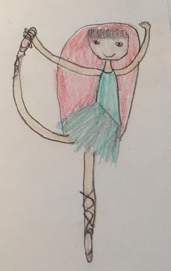
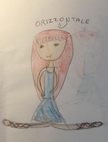
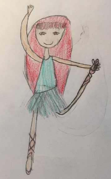
· The repetition of the movement. It has as a result, according to Pavlov, the formation of new central integrations6 establish (corticalizzazione).
· Spatial summation stimuli: the simultaneous activation of several excitatory synapses on the same neuron leads to the achievement of the current threshold that allows the generation of an action potential by the neuron, and therefore the propagation of the stimulus. From the point of view of practical application, this means for us that administering multiple stimuli at the same time allows us to enhance the motor response by increasing the number of motor neurons involved. To this we must add that the intensity of the stimulus increases the muscle response, therefore even a modulation of this intensity is one of the tools available to us to refine the effectiveness of the proposed exercises.
· Time summation stimuli: repeated stimulations increase the motor response.
· Type of contraction. Isometric contractions, concentric isotonic and eccentric isotonic: the first are contractions without moving the joint heads, the second with approach and the last with removal of the joint heads. The first case occurs, for example, when we keep a heavy object raised without moving it. The second is when we lift the shopping bag off the ground, the third when we rest it on the ground. These three types of contractions
In 1987 an Italian physiotherapist, Giuseppe Monari, after taking a PNF course in California, introduced the metodo Kabat in Italy,.
The use of this method led him over the years to introduce his own elaborations which led to the creation of a much more complex and articulated approach than the original model, so much so as to justify and make a change of name necessary to distinguish those that have in fact become two different modes of intervention, while respecting the initial formulations that report to Kabat and his working group.
Here we will retrace the steps that led to the current RMP-Elaboration of the Kabat Concept, taking as a reference the story of this evolution contained in the introduction to the edition of 2014 of “Progressive Modular Rebalancing, Elaboration of the Kabat concept7”In G. Monari.
Monari tells us about a first evolution that dates back to the years 1974-1980 in which the importance of biarticularity and complex monoarticularity was identified.
A biarticular muscle it is a muscle that includes two joints between its joint heads and consequently its action can have an effect on both. The rectus femoris, for example, can act as a hip flexor and / or as a knee extensor. Working in biarticularity for the rectus femoris means acting on both joints and therefore dividing its work on both its insertion heads in an appropriate percentage according to the specific need of the motor act to be performed. Monari calls this way of working the muscle "intelligent function".
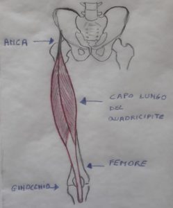
Having to divide its strength both upstream and downstream with respect to its length, the rectus femoris forces other muscles to come to its aid to perform both functions in the best possible way, of hip flexion (asking for help from the psoas muscle) and knee extension (asking for help from the vast muscles) and this involvement will also improve the performance of the supporting muscles over time.
The biarticular work forces the rectum to vary greatly its length and in this way improves its elasticity. If the rectum is exercised separately only as a hip flexor or only as a knee extensor, it is not forced to intelligently divide its work and this requires cortical involvement (cerebral) of lower quality level and determines a lower neuroplasticity effect. The difference in level of cortical involvement was demonstrated by functional transcranial color doppler. Furthermore, by working only on one of his adverts, he does not need any particular length variations because the one he needs is obtained by stretching at the level of the other advert. In bi-articular work, the muscle starts from a maximum shortening to reach a maximum elongation.
Per complex monoarticularity it is meant that a monoarticular muscle works simultaneously in all three of its functions: flexion-extension, abduction-adduction, rotations. In this way it will be able to reach the state of maximum shortening. As well as the work in biarticularity, even the use of complex monoarticularity involves greater cortical involvement and therefore greater neuroplasticity.
A second evolution of the method then took place between 1974 and the 1980, when it was identified in postural steps the key to understanding the recruitment capacity of the trunk in its four rotation functions, flexion, extension and inclinations. The postural passages thus become a refined tool for evaluating the functions of the trunk, but at the same time they also constitute a basic platform on which to structure the therapeutic exercises to be proposed to rehabilitate those components of the trunk that are deficient.
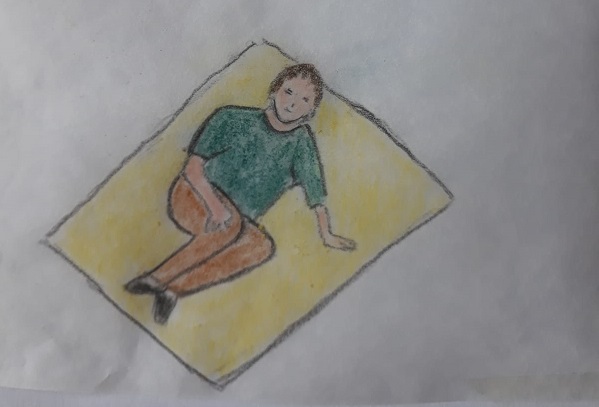
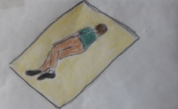
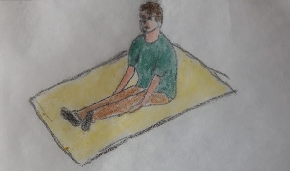
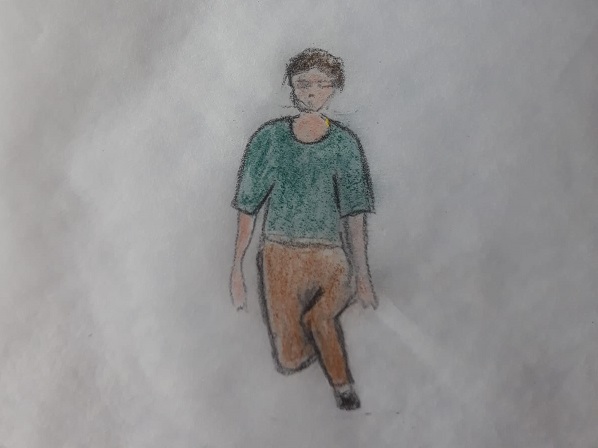
Between 1978 and the 1986 there was the introduction of pyramid progressions to evaluate and intervene specifically on the problems of Balance who, as known, is given by the ratio between the width of the support base and the height from the ground of the center of gravity of a body.
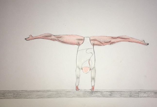
The balance, like any of our other skills, it is actually the result of training-learning, more or less aware (the child “trains” from birth and for more than a year to achieve the necessary balance for an upright position), which involves changes in our central nervous system. This is possible thanks to the neuroplasticity of our nervous system, or the ability to change in response to stimuli.
Working on balance means proposing to the patient exercises set on a targeted variation of these two parameters that gradually allows the patient to reach an upright position (where the support base is minimal and the center of gravity at the maximum height from the ground) having filled those gaps that made it impossible to achieve or that made it precarious. By pyramidal progression we mean all that progression of postural passages that allow us to pass from the supine position (maximum support base and minimum height of the center of gravity) to standing position (or from the prone position, or from the lying position on the side). The evaluation of how the patient deals with these pyramidal progressions allows to intercept the moment in which deficiencies begin to appear and therefore to identify the level of exercises to be proposed and also allows to evaluate whether the patient is able to reach the upright position and the walking. This work was fundamental because it made it possible to overcome a consolidated modus operandi which was to directly exercise the lacking function to improve it.: if the patient walks badly, let him walk and by exercising he will improve. These studies suggest the fact that it is probably more appropriate to identify the deficiencies upstream of the impaired function and rehabilitate them in a specific way in order to properly re-establish the function. (for example walking).
Subsequently, the importance of muscle elasticity was highlighted for a muscle to best exert its contractile power. A muscle that has undergone shortening is a muscle that has lost elasticity. The greater the ability of a muscle to reach maximum elongation and maximum shortening, the greater its contractile capacity. Furthermore, a shortened muscle limits the action of its antagonist because it acts as a brake (when a muscle contracts its antagonist must relax and stretch). Muscle shortening would also have an effect on the sensitivity of the muscle spindles which would increase the excitability of the stretch reflex.
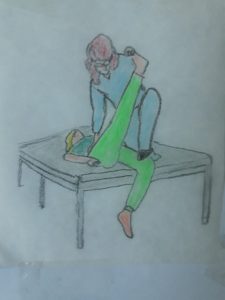
Hence the pivotal role that is attributed by the RMP to the achievement and maintenance of muscle lengths physiological in every area, but in particular in that of neurological pathologies, given that an alteration is found in these patients, which facilitates the establishment of pathological patterns and can negatively affect shoulder pain which can often be encountered in particular by the hemiplegic patient: "..The therapist even before dealing with" muscle strengthening "must worry about" muscle lengths "". Therefore, a "real" method of evaluating muscle lengths was prepared, that ensures effective control of the position of the joint heads during the evaluation, preventing the escape routes of the system that we call "compensation".
These are the main innovations that are indicated in the writing of G. Monari to whom I refer here. They are not the only ones but I believe they are sufficient to give an idea of the theoretical basis of this method and its rehabilitation potential.
___________________________________________________________
1Noel-Ducret F. Metodo in Kabat. Proprioceptive neuromuscular facilitation. Encycl Med Chir (Scientific and Medical Editions Elsevier SAS Paris), Rehabilitation Medicine, 26-060-C-10,2001, 18 p.
E. A lot of. The kabat method. Proprioceptive neuromuscular facilitation. Publisher Marrapese. 1997.
G. Monari. Progressive Modular Replenishment. Elaboration of the Kabat concept. Edi-ermes, 2004.
1aNG. Monari. Progressive Modular Rebalancing. Elaboration of the Kabat concept. Edi-ermes. 2013
2G. Monari. Progressive Modular Replenishment. Elaboration of the Kabat concept. Edi-ermes, 2004.Lots. Use of proprioceptive neuromuscular techniques for reeducation and education of the sporting gesture. Switzerland Zeits Sport Medb 1985; :00-104
3 E. A lot of. The Kabat method. Proprioceptive neuromuscular facilitation. Publisher Marrapese. Roma. 1997
4 place.
5 Ibidem pag 113
6place
7G. Monari. Progressive Modular Rebalancing. Elaboration of the Kabat concept. Edi-ermes. 2013