Pelvic floor and abdomen in women
The pelvic floor, also known as the pelvic diaphragm, it is the support base of the pelvic viscera (bladder, straight, pelvic genital organs and terminal part of the urethra) contained in the small pelvis, also called pelvis. It is a structure made up of muscles, fascial system and ligaments that close the lower part of the abdomen.
In women it has two orifices that communicate with the outside respectively, in a posterior anterior direction, intestine, uterus and bladder:
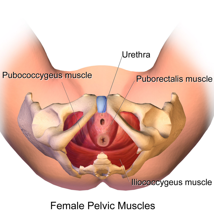
- Rectal hiatus – a centrally positioned slot, which allows the passage of the anal canal.
- Urogenital hiatus – a fissure located anteriorly, which allows the passage of the urethra and vagina
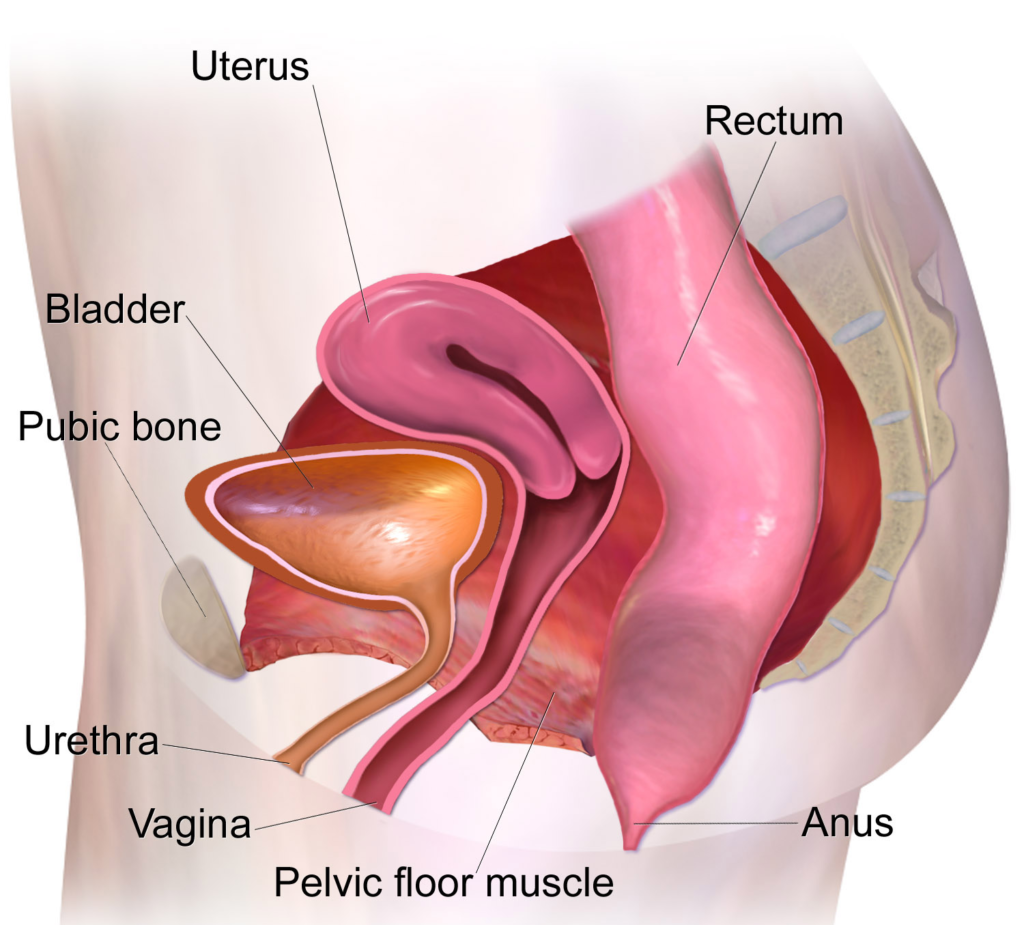
The function of the pelvic floor is to ensure stability and support to the pelvic organs, avoiding their prolapse and to offer adequate support to the normal rhythms of continence and bladder and rectal emptying thanks to a refined management of the possibilities of contraction and relaxation of its various muscles and elasticity and resistance of its muscular structures, fascial and ligament.
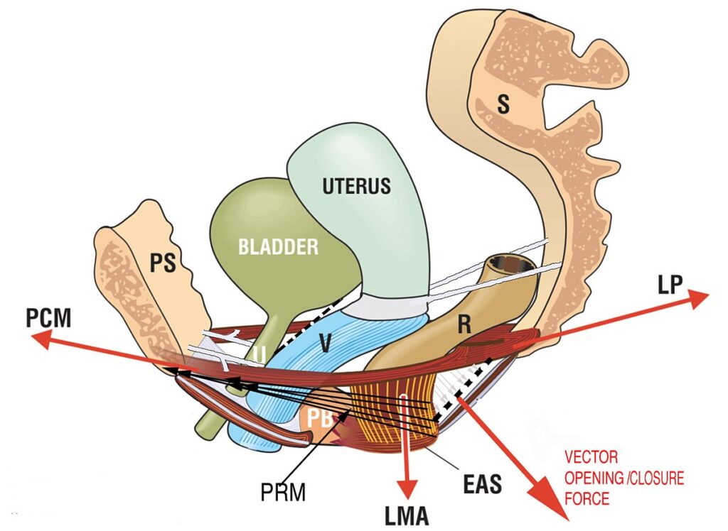
Four major vector muscle forces work in a coordinated manner to close or open the urethral exit conduits “U”, vagina “V” and straight “R”. It is evident that the arrows contract against the anterior and posterior suspensory ligaments (the PUL, connected to the urethra; l’ USL, connected to the uterus).
Each muscle layer of the pelvic floor performs specific functions:
Lo deeper muscle layer, also called pelvic diaphragm, it is occupied by the levator ani muscle, responsible for managing restraint and intestinal evacuation. It is a bilateral muscle made up of three heads for each hemilate: ilio-coccygeus, ischio-coccygeal and pubo-coccygeal.
The deep transversus perineum muscle and the puborethral ligaments constitute it intermediate layer also called urogenital diaphragm which has the function of supporting the urethra, the uterus and vagina;
Lastly there is it superficial layer which houses the sphincters, made up of four muscles:
- constrictor of the vagina
- superficial transverse perineum
- sphincter of the anus.
These are muscles that must constantly modulate their contraction and relaxation activity in an extremely fine way to respond to our postural changes, to the different activities we carry out during the day to the different environmental situations. Most of their activity takes place without us realizing it or paying attention to it and in any case we are normally not able to perceive the activation of these different muscles or even of the different muscle planes.
For the pelvic floor to best perform its functions, the muscles that make it up must be in good health. One of the objectives of pelvic floor rehabilitation is to restore the correct lengths when they have undergone shortening, remove any contractures and restore tone if hypotonic.
Atnmain problems related to the pelvic floor:
- Urinary incontinence
- Difficulty emptying the bladder
- Anal incontinence.
- Constipation
- Sags and prolapses
- Chronic pelvic pain
- Genital and lower urinary tract pain
Abdominal diastasis and pelvic floor
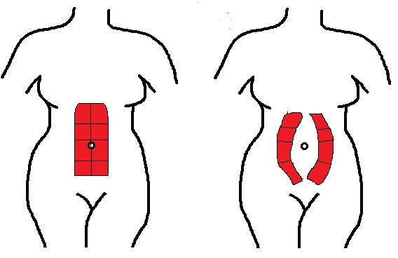
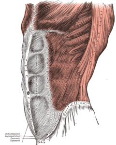
To carry out its functions correctly, the pelvic floor needs to work in synergy with valid abdominal muscles. In some cases, however, it happens that, after a pregnancy, a problem with the rectus abdominis muscles occurs. Under normal conditions the rectus muscle of the right side of the body is adjacent to the rectus muscle of the left side but pregnancy, due to the increase in the volume of the abdomen, may involve their separation with a more or less wide spacing. This phenomenon is called diastasis of the rectus abdominis muscles. This condition negatively affects the validity of the pelvic floor.
The diastasis in most cases can be treated conservatively with hypopressive gymnastics thanks to which it is possible to obtain a reduction of the amplitude e, in some cases, its complete resolution.
