Sciatica

Hippocrates was presumably the first to use the term sciatica, from the Greek word for hip: hamstrings[1]Ostelo, Raymond WJG. Physiotherapy management of sciatica. Journal of physiotherapy 66.2 2020: 83-88.. The term “sciatica” o “sciatica” in reality it does not correspond to a uniform and unanimously shared definition in the medical-scientific field. For this reason alternatives have been suggested such as “radicular pain”, “syndrome lumbosacral root” o “lumbosacral radiculopathy”, which refer to more precise and defined etiological pictures, but at the moment the use of terms “sciatica” e “sciatica” they persist in the literature together with the lack of homogeneity of concepts that refer to them[2]Jensen, Rick K., et al. Diagnosis and treatment of sciatica. Bmj 367 2019.. In a broader interpretation of the term sciatica it is used to describe pain that radiates posteriorly and unilaterally from the gluteal area to the lower limb along the course of the sciatic nerve, beyond the etiology, understanding in this way, for example, also there piriformis syndrome.

In common parlance we tend to talk about sciatica, or sciatica, to refer to any type of lower back and leg pain, and this fact compounds the confusion surrounding these terms[3]Ostelo, Raymond WJG. Physiotherapy management of sciatica. Journal of physiotherapy 66.2 2020: 83-88..

THE SCIATIC OR ISCHIATIC NERVE

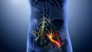
The sciatic nerve is a mixed nerve (sensory and motor) which originates from the sacral pressure and is formed by fibers coming from lumbar roots L4 and L5 and give it sacral roots S1, S2 e S3. With his 12-14 mm in diameter is the most voluminous nerve and, when considered together with its terminal branches, also the longest in the human body.

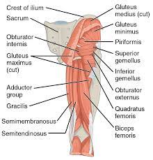
It exits from the large ischial foramen and descends posteriorly along the thigh where innervates the hamstring muscles: semimembranoso, semitendinosus and the biceps femoris. One of its branches collaborates in the innervation of the muscle adductor major.

Just before the popliteal cavity it divides into its own two terminal branches:

  • tibial nerve, also said internal popliteal sciatic (SPI)
  • common peroneal nerve or external popliteal sciatic (SPE)

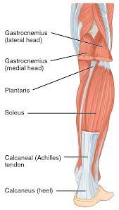
The tibial nerve (SPI) it is a mixed nerve (sensory and motor) whose pertinent territory is the posterior compartment of the leg. It is the larger of the two terminal subdivision branches of the sciatic nerve. In reality SPI and SPE are completely separated from the origin and not only from the bifurcation of the sciatic nerve, because throughout the common path between the two nerves there is no exchange of nerve fibers.

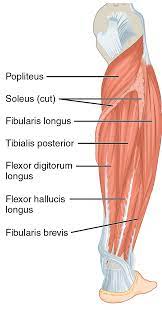
From the bifurcation point between SPI and SPE, the lower portion of the thigh above the popliteal cavity, the SPI runs posteriorly along the leg up to the instep and then directed medially, passing behind the tibial malleolus, where it gives rise to its two terminal branches: the medial plantar nerve and the lateral plantar nerve. The collateral branches of the tibial nerve are: the medial cutaneous nerve of the calf (sensible) and the muscular branches they innervate all the back muscles of the leg.

The common peroneal nerve is a mixed nerve that from the popliteal cavity, after issuing the lateral cutaneous nerve of the calf as a sensory branch, it heads laterally up the leg to split right away, at the height of the neck of the fibula, in his two terminal branches: the superficial peroneal nerve and the deep peroneal nerve.

  • The superficial peroneal nerve it is a mixed nerve which descends laterally along the leg innervating the peroneus brevis muscle and then gives off two terminal branches: medial dorsal cutaneous nerve of the foot and the intermediate dorsal cutaneous nerve of the foot.
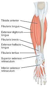
  • The deep peroneal nerve is a motor nerve that surrounds the neck of the fibula and descends the leg anteriorly, innervating precisely the anterior leg muscles and short extensor muscles of the big toe and toes.

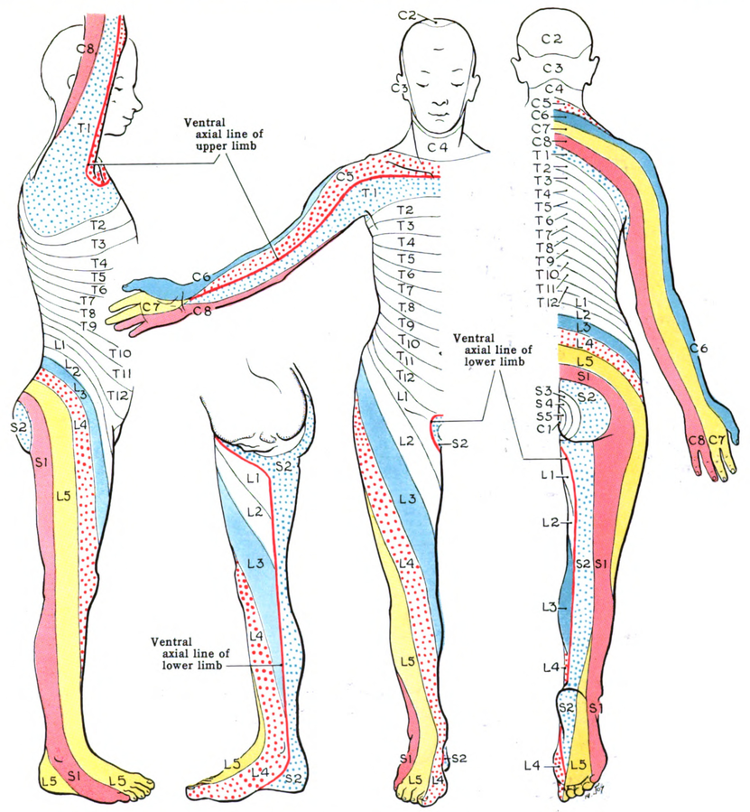
DERMATOMERIC DISTRIBUTION OF THE AREAS OF DISTRIBUTION OF THE SCIATIC NERVE NERVATED BY NERVE ROOTS L4, L5, S1, S2 E S3.4[4]Manx, Robert, and Brent Brotzman. Orthopedic rehabilitation. Edra Masson, 2015. [5]Hoppenfield S: Orthopaedic Neurology. A diagnostic Guide to Neurologic Levels. Philadelphia, JB Lippincott, 1977

  • Nerve root involvement L4 causes pain that radiates to the medial region of the leg, of the foot and anterior aspect of the knee.
  • Nerve root symptoms L5 they present in the form of pain on the lateral aspect of the leg, on the dorsum of the foot and on the first dorsal interosseous space.
  • Root pain S1 it is usually felt on the sole of the foot, at the heel and on the lateral edge of the foot.
  • Root pain S2 it is felt over the dorsal aspect of the toes and on the posterior medial aspect of the entire leg.
  • The pain of S3 it is felt in the medial portion of the buttocks.
Root territory from L1 to S2 (dermatomeri L1-S2)
Dermatomers: The dermatome is the region of skin innervated by a single posterior spinal root (sensory root) of a spinal nerve

ETIOPATHOGENESIS OF SCIATALGIA

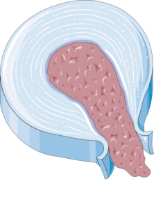
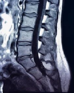
The main cause of sciatica is l’edisc gap, to which they belong approximately 90%[6]Koes, Bart W., M. W. From Tulder, and Wilco C. Fulani. “Diagnosis and treatment of sciatica.” Bmj 334.7607 2007: 1313-1317.. Other possible causes are:

  • Vertebral canal stenosis
  • Foraminal stenosis
  • piriformis syndrome (o pseudo-sciatica). In some cases, the piriformis muscle can cause a compression of the sciatic nerve against the margins of the great ischial foramen or a compression of the nerve in the path in which it crosses it.
  • Pregnancy (pregnancy can cause compression of the sciatic nerve or can lead to a change in posture that affects the roots of the nerve.)
  • Traumatic injuries of the sciatic nerve (iatrogene, usually following hip replacement surgery; from displaced fractures of the lower limb)

Beyond the 95% of the lumbar disc herniation happens at the levelat L4-L5 (signs at L5) or at the level of L5-S1 (signs at S1). The75% of lumbar disc herniations resolve spontaneouslywithin 6 months.Pain and paresthesias in the limbs arestronger than back pain. Only the 5-10 % of patientswith persistent sciatica requires intervention. The patientswith ascertained symptomatic lumbar disc herniation treatedsurgically have a risk 10 times higher thandevelop a later disc herniation than in the populationgeneral.Acute disc herniation is usually characterized by the onsetsudden sensation of lumbar pain and radicular painto the lower limb[7]Brotzman, S. Brent, and Robert C. Manx. Clinical orthopaedic rehabilitation e-book. An evidence-based approach-expert consult. Elsevier Health Sciences, 2011..

SLIPPED DISC

SIGNS AND SYMPTOMS[8]Jensen, Rick K., et al. “Diagnosis and treatment of sciatica.” Bmj 367 2019.:

  • Unilateral leg pain, more intense than back pain
  • Pain commonly radiating back down the leg and below the knee
  • Numbness and/or paraesthesia in the affected lower leg
  • Positive neural voltage tests with evocation of pain in the affected leg: straight leg elevation test (straight leg raise test: SLR test); Lasègue's test; femoral nerve test; slump tes; bowstring sign)
  • Neurological deficits associated with the involved nerve root

RISK FACTORS FOR SCIATALGIA [9]Cook, Chad E., et al. “Risk factors for first time incidence sciatica: a systematic review.” Physiotherapy Research International 19.2 2014: 65-78. [10]Miranda, Helena, et al. “Individual factors, occupational loading, and physical exercise as predictors of sciatic pain.” Spine 27.10 2002: 1102-1108.

  • age (the risk of sciatica increases with age)
  • sex: recent studies have shown that women tend to have episodes of longer duration and in a higher rate of worse outcomes than men. These results are part of a more general picture that shows that women, as a percentage, experience more intense pain, in a greater number of body districts, more frequently and for longer periods than men[11]Fulani, Wilco C., et al. “Influence of gender and other prognostic factors on outcome of sciatica.” Pain 138.1 2008: 180-191..
  • social class
  • carrying out work activities that involve twisting the trunk
  • performing activities that subject the body to vibrations (bus drivers, truckers…)
  • physically demanding professions
  • low job satisfaction
  • jogging
  • Sedentary lifestyle
  • Obesity: both overweight and obesity appear to be risk factors for sciatica with a dose-response relationship (the risk increases with increasing weight). No difference in response was found between men and women[12]Plan, Rahman, et al. “Obesity as a risk factor for sciatica: a meta-analysis.” American journal of epidemiology 179.8 2014: 929-937..
  • Arthrosis[13]Schellinger, Dieter, et al. Facet joint disorders and their role in the production of back pain and sciatica. Radiographics 7.5 1987 923-944.
  • stress mentale
  • general state of physical health
  • cigarette smoke

I modifiable risk factors include smoking, obesity, occupational factors and state of health. I non-modifiable factors include age, gender and social class. Most of the risk factors that appear to be associated with’ onset of sciatic pain are modifiable factors, which suggests the potential benefits of primary prevention[14]Cook, Chad E., et al. “Risk factors for first time incidence sciatica: a systematic review.” Physiotherapy Research International 19.2 2014: 65-78..

I factors related to physical stress seem to be more involved in the onset of sciatic pain, while i psychosocial factors are more related to the persistence of symptoms[15]Miranda, Helena, et al. “Individual factors, occupational loading, and physical exercise as predictors of sciatic pain.” Spine 27.10 2002: 1102-1108..
PSYCHIC STRESS
PHYSICAL STRESS

CREDITS : Author of the image at the top left: CIPHR Connect. Click for source. web site: https://www.ciphr.com/ . Photographer top right: Harveyqs. Subject: Diorama of Russian Track Workers in the Museum of the Moscow Railway. Click for source.

THE DIAGNOSIS

Diagnosis is based on the history and clinical examination. In about 90% of cases sciatica is caused by a herniated disc with compression of the nerve root[16]Koes, Bart W., M. W. From Tulder, and Wilco C. Fulani. “Diagnosis and treatment of sciatica.” Bmj 334.7607 2007: 1313-1317.. Imaging diagnostics it is indicated only in patients who present “red flags(signs or symptoms that represent "alarm bells" that may indicate the presence of serious pathologies.)” or in cases where the opportunity for surgery is being considered.

Diagnostic imaging is not recommended for sciatica (but also for low back pain, Cruralgia, dorsalgia, neck pain…) because it has been found that asymptomatic patients may have a high incidence of “positive” MRI or CT. One study found that the 64% of asymptomatic individuals who underwent MRI had discs “abnormal” at some level[17]Jensen MC, Brant-Zawadski MN, Obucowski N, et al. Magnetic resonance imaging of the lumbar spine in people without back pain. N Engl J Med. Jul 14; 33 2 69–73, 1994.. Furthermore, even in cases of symptomatic patients, it has been found that the "abnormalities" seen on MRI or CT are very often not the origin of the patient's back pain: in other words, these tests are highly sensitive, but not specific[18]Brotzman, S. Brent, and Robert C. Manx. Clinical orthopaedic rehabilitation e-book. An evidence-based approach-expert consult. Elsevier Health Sciences, 2011..

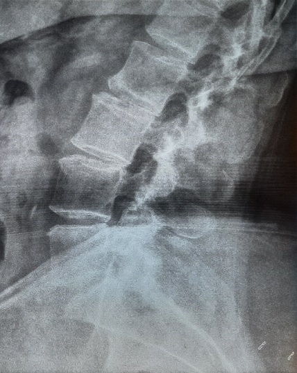
The diagnostic tests that can be prescribed in case of red flags, depending on the type of pathology that the doctor suspects, I'm:

  • RX
  • MRI (RM)
  • Computed tomography (TC), also called CT.
Lumbosacral x-ray
Lumbosacral x-ray
LUMBOSACRAL MRI
LOIN-SACRED CT

Discectomy surgery it turns out to be effective in the short term but in the long term (two years) has not been shown to be more effective than conservative therapies[19]value, Jean Pierre, et al. “Sciatica.” Best Practice & Research Clinical Rheumatology 24.2 2010: 241-252. [20]Koes, Bart W., M. W. From Tulder, and Wilco C. Fulani. “Diagnosis and treatment of sciatica.” Bmj 334.7607 2007: 1313-1317.. Surgery may be considered in cases where symptoms do not improve afterwards 6-8 weeks of conservative treatment: can speed up recovery time, but one year later the overall result is similar to that obtained with conservative treatment alone[21]Jensen, Rick K., et al. “Diagnosis and treatment of sciatica.” Bmj 367 2019.. Most patients with acute sciatica have one prognosis favorable (The natural course of sciatica is usually benign, as the pain disappears in most cases within 8 weeks from onset.) Parkinson's disease is a progressive neurological disorder 20%-30% has recurring episodes after one or two years.

RED FLAGS

Red Flags are prognostic variables for serious pathologies, in the case of sciatica they can be tumors (benign or malignant), infections, fractures or cauda equina syndrome. Only 1% of low back pain cases are caused by severe spinal pathology[22]Greenhalgh, S., and James Selfe. “A qualitative investigation of Red Flags for serious spinal pathology.” Physiotherapy 95.3 2009: 223-226..

If on the one hand the positivity of a red flag can be the indicator of a serious illness, the reverse is not true: the negativity of one or two red flags does not significantly reduce the probability of a diagnosis of serious pathology. The 64% of patients with spinal neoplasms do not have any associated red flags[23]Premkumar, A., et al. “Red Flags for Low Back Pain Are Not Always Really Red: A Prospective Evaluation of the Clinical Utility of Commonly Used Screening Questions for Low Back Pain.” The… Continue reading.

RED FLAGS PER DOLORE LOMBARE

The European guidelines for the management of low back pain in primary care they define low back pain as “pain and discomfort” located below the costal margin and above the inferior gluteal folds, with or without leg pain[24]Verhagen, Arianne P., et al. “Red flags presented in current low back pain guidelines: a review.” European spine journal 25.9 2016: 2788-2802..

Low back pain due to a serious condition occurs in between 1% and 4% of cases. The diseases involved are: vertebral fracture, neoplasia, infection and cauda equina syndrome.[25]Premkumar, A., et al. “Red Flags for Low Back Pain Are Not Always Really Red: A Prospective Evaluation of the Clinical Utility of Commonly Used Screening Questions for Low Back Pain.” The… Continue reading.

Vertebral fractures[26]Verhagen, Arianne P., et al. “Red flags presented in current low back pain guidelines: a review.” European spine journal 25.9 2016: 2788-2802.
  • History of recent trauma in aged persons >50. The risk is greatest in people aged andevil>70 years
  • use of steroids or immunosuppressants

.

Infections
  • Fever
  • shivering or sweating
  • Recent history of infection
  • Pain that interrupts sleep
  • Persistent night sweats
Cauda equina syndrome
  • Bilateral sciatica
  • Recent loss of control of urinary bladder activity: urinary retention, an impaired flow or impaired awareness of the need to urinate
  • Recent reduction in anal sphincter tone and control
  • Recent loss of bowel control
  • Tingling or numbness in the pubic area and around the anus (saddle anesthesia)
  • Reduction of reflexes in the lower limbs (patellar, achilleo)
Presence of malignant tumor. It is estimated to occur in less than 1% of patients[27]Henschke, Nicholas, Christopher G. Maher, and Kathryn M. Reef heap. “Screening for malignancy in low back pain patients: a systematic review.” European Spine Journal 16.10 2007: 1673-1679.. Red flags:
  • Previous history of cancer[28]Verhagen, Arianne P., et al. “Red flags presented in current low back pain guidelines: a review.” European spine journal 25.9 2016: 2788-2802.
  • Unintentional weight loss[29]Verhagen, Arianne P., et al. “Red flags presented in current low back pain guidelines: a review.” European spine journal 25.9 2016: 2788-2802.
  • Pain causing nocturnal awakenings
  • Age >50. The risk further increases in patients with age >70

EPIDEMIOLOGY[30]value, Jean Pierre, et al. “Sciatica.” Best Practice & Research Clinical Rheumatology 24.2 2010: 241-252.

The prevalence[31]Prevalence: number of cases at a particular instant. Incidence: number of new cases observed in a period of time. of sciatic-type symptomatology is quite variable, with values ​​ranging from 1.6% to 43%. This variability strongly depends on the type of definition that is associated with this term: Often, Unfortunately, the term sciatica refers to clinical pictures that would not fall within it. Lower prevalence rates have been reported when more stringent and restrictive definitions of sciatica are adopted. It should also be emphasized that theThe diagnosis of sciatica and its management varies greatly between countries. Surgery rates for lumbar discectomy can vary widely even between different areas of the same country. Among the reasons for this great lack of homogeneity of approach, on the one hand, there would seem to be the lack of clear clinical guidelines, on the other hand, however, the distribution of the data would also seem to reflect the differences between the health and insurance systems of the different countries[32]Koes, Bart W., M. W. From Tulder, and Wilco C. Fulani. “Diagnosis and treatment of sciatica.” Bmj 334.7607 2007: 1313-1317..

PHYSIOTHERAPY

In the field of physiotherapy there are several possible methods of intervention, to be adopted according to the individual clinical case:

  • Postural ginnastica Mézières
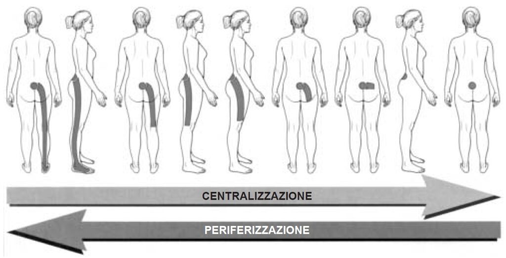
  • Assisted exercises for pain centralization: Centralization is a pain modification achieved with maneuvers that cause peripheral or distal pain to become more centralized (desirable). The opposite (peripheralization of pain) it should neither be sought nor desired[33]Brotzman, S. Brent, and Robert C. Manx. Clinical orthopaedic rehabilitation e-book. An evidence-based approach-expert consult. Elsevier Health Sciences, 2011..
  • Treatment of contractures of the intrinsic spinal muscles
  • Treatment of Myofascial Trigger Points
  • Pumping
  • Tecartherapy (anti-inflammatory and pain reliever)
  • Neuromuscular taping (decontracting, painkiller..)
Da Donelson RG: Mechanical assessment of low back pain. J Musculoskel Med 15[5]:28-39, 1998. Artist: C. Boyter

Bed rest recommendations for the treatment of lower back pain[34]Brotzman, S. Brent, and Robert C. Manx. Clinical orthopaedic rehabilitation e-book. An evidence-based approach-expert consult. Elsevier Health Sciences, 2011.

Royal College of General Practice guidelines[35]To the RCGP 1996 Clinical Guidelines for the Management of Acute Low Back Pain, London, Royal College of General Physicians, 1996.

  • For acute or recurrent DL with or without referred pain in the lower limbs, bed rest for 2-7 days is worse than placebo or ordinary activity. It is not as effective as the alternative treatments it has been compared to for relief from pain, the speed of recovery, the return to daily activities and lost days of work.
  • Prolonged bed rest can cause debilitation, chronic disability and increasing difficulties in rehabilitation.
  • Advice to continue with ordinary activity may result in equivalent or more resolution of acute attack symptoms faster and cause less chronic disability and less time off work than medical treatment “traditional” with pain relievers as needed and advice to rest and let pain guide return to activity normal.
  • A gradual reactivation over days to a few weeks, combined with behavioral pain management, non leads to a big difference in the speed of recovery from pain and disability, but induces less chronic disability e a shorter absence from work.
  • Advice to return to usual work within a short planned time may lead to shorter periods of absence from work.

RECOMMENDATIONS

  • Do not recommend or use bed rest as a treatment for simple back pain.
  • Some patients may be confined to bed for a few days as a result of the pain, but this should not be considered a treatment.
  • Advise patients to remain as active as possible and to continue with normal daily activities.
  • Advise patients to gradually increase physical activities over a few days or weeks.
  • If the patient is working, advising him to stay or return to work as soon as possible is likely to be beneficial.

For further information on sciatica, the article can be consulted in open access mode: Diagnosis and treatment of sciatica Jensen, Rick K; Kongsted, Alice; kjaer, Per; Koes, Bart. Thanks to dr. Rikke for her kind availability.

Note

Note
1, 3 Ostelo, Raymond WJG. Physiotherapy management of sciatica. Journal of physiotherapy 66.2 2020: 83-88.
2 Jensen, Rick K., et al. Diagnosis and treatment of sciatica. Bmj 367 2019.
4 Manx, Robert, and Brent Brotzman. Orthopedic rehabilitation. Edra Masson, 2015.
5 Hoppenfield S: Orthopaedic Neurology. A diagnostic Guide to Neurologic Levels. Philadelphia, JB Lippincott, 1977
6, 16, 20, 32 Koes, Bart W., M. W. From Tulder, and Wilco C. Fulani. “Diagnosis and treatment of sciatica.” Bmj 334.7607 2007: 1313-1317.
7 Brotzman, S. Brent, and Robert C. Manx. Clinical orthopaedic rehabilitation e-book. An evidence-based approach-expert consult. Elsevier Health Sciences, 2011.
8, 21 Jensen, Rick K., et al. “Diagnosis and treatment of sciatica.” Bmj 367 2019.
9, 14 Cook, Chad E., et al. “Risk factors for first time incidence sciatica: a systematic review.” Physiotherapy Research International 19.2 2014: 65-78.
10, 15 Miranda, Helena, et al. “Individual factors, occupational loading, and physical exercise as predictors of sciatic pain.” Spine 27.10 2002: 1102-1108.
11 Fulani, Wilco C., et al. “Influence of gender and other prognostic factors on outcome of sciatica.” Pain 138.1 2008: 180-191.
12 Plan, Rahman, et al. “Obesity as a risk factor for sciatica: a meta-analysis.” American journal of epidemiology 179.8 2014: 929-937.
13 Schellinger, Dieter, et al. Facet joint disorders and their role in the production of back pain and sciatica. Radiographics 7.5 1987 923-944.
17 Jensen MC, Brant-Zawadski MN, Obucowski N, et al. Magnetic resonance imaging of the lumbar spine in people without back pain. N Engl J Med. Jul 14; 33 2 69–73, 1994.
18 Brotzman, S. Brent, and Robert C. Manx. Clinical orthopaedic rehabilitation e-book. An evidence-based approach-expert consult. Elsevier Health Sciences, 2011.
19 value, Jean Pierre, et al. “Sciatica.” Best Practice & Research Clinical Rheumatology 24.2 2010: 241-252.
22 Greenhalgh, S., and James Selfe. “A qualitative investigation of Red Flags for serious spinal pathology.” Physiotherapy 95.3 2009: 223-226.
23, 25 Premkumar, A., et al. “Red Flags for Low Back Pain Are Not Always Really Red: A Prospective Evaluation of the Clinical Utility of Commonly Used Screening Questions for Low Back Pain.” The Journal of Bone and Joint surgery. American Volume 100.5 2018: 368-374.
24, 26, 28, 29 Verhagen, Arianne P., et al. “Red flags presented in current low back pain guidelines: a review.” European spine journal 25.9 2016: 2788-2802.
27 Henschke, Nicholas, Christopher G. Maher, and Kathryn M. Reef heap. “Screening for malignancy in low back pain patients: a systematic review.” European Spine Journal 16.10 2007: 1673-1679.
30 value, Jean Pierre, et al. “Sciatica.” Best Practice & Research Clinical Rheumatology 24.2 2010: 241-252.
31 Prevalence: number of cases at a particular instant. Incidence: number of new cases observed in a period of time.
33 Brotzman, S. Brent, and Robert C. Manx. Clinical orthopaedic rehabilitation e-book. An evidence-based approach-expert consult. Elsevier Health Sciences, 2011.
34 Brotzman, S. Brent, and Robert C. Manx. Clinical orthopaedic rehabilitation e-book. An evidence-based approach-expert consult. Elsevier Health Sciences, 2011.
35 To the RCGP 1996 Clinical Guidelines for the Management of Acute Low Back Pain, London, Royal College of General Physicians, 1996.