Scoliosis

The word "scoliosis" comes from the Greek “scoliosis”, which means bowing (“scoliosis” smeans "wrong") and its first use is attributed to Galen of Pergamum, Greek physician of the 2nd century, to talk about pathological deviations of the spine.

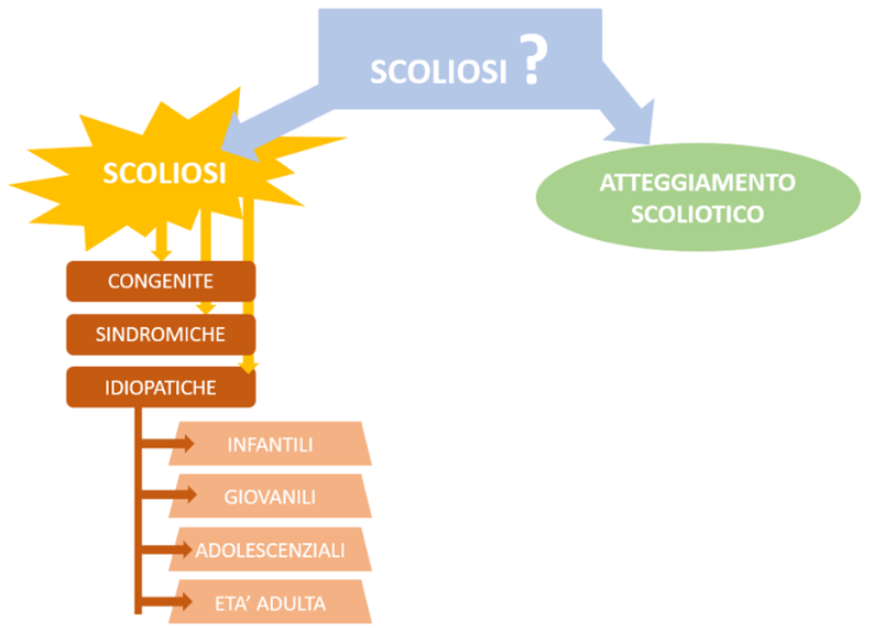
The first big distinction to make when it comes to scoliosis is between real scoliosis and pseudo-scoliosis or scoliotic attitude..

In the scoliotic or pseudo-scoliosis attitude, only the lateral inclination of the column is present. There is no vertebral rotation associated with the inclination and the hump is absent. In the supine position the inclination is completely reducible, that is, it disappears.

The scoliotic attitude may be due to incorrect postural habits (which can be linked, for example, to a lack of muscles, to an analgesic response to visceral or musculoskeletal problems, a fascial disturbance, ..). In some cases the underlying cause is a difference in length between the lower limbs. The peculiarity of these pseudo-scoliosis is that it just comes resolved the cause, the pseudo-scoliosis disappears.

The scoliotic attitude it does not involve deformation of the vertebral bodies.

The picture changes completely when it comes to real scoliosis. The commonly adopted definition tells us that:

"Scoliosis is a deformation of the Spine in the three planes of space with an angle greater than 10 ° on the frontal plane[1]Terminology Committee of the Scoliosis Research Society. A glossary of terms. Spine1976;1:57-8.”.

Scoliosis, unlike the scoliotic attitude, present, associated with the lateral inclination of the column, also there vertebral rotation that, at the dorsal level, includes the troupe of gibbon. The dorsal vertebrae, indeed, articulate with the rib cage. When a group of vertebrae goes towards rotation, it also carries with it the coasts connected to it thus disharmonizing the coherence of the structure of the rib cage. The vertebral body of the involved vertebrae rotates towards the opposite side with respect to the inclination: if the column, as in the example shown in the image, is tilted to the left (left concavity), the vertebral body rotates to the right. The spinous process of the vertebrae moves to the concave side. The hump is formed on the convex side.

The deviations of the column, in scoliosis, remain also in the supine position. This type of scoliosis involves structural modifications at the bone level (mainly of vertebrae and ribs) but also at the level of soft tissues such as fascial structures, ligamentous and muscular.

CLASSIFICATION OF SCOLIOSIS

Scoliosis is classified, based on etiology, in three different macro categories[2]Coillard, Christine, Alin B. Circus, and Charles H. reward. “SpineCor treatment for Juvenile Idiopathic Scoliosis: SOSORT award 2010 winner.” Scoliosis 5.1 2010: 1-7.:

· CONGENITAL (associated with an abnormal formation of the vertebrae)

· SYNDROME (associated with problems in the neuromuscular systems, bone or connective tissue.). They can be divided into two macro categories:

o Neuromuscular scoliosis. They are caused by the insufficiency of active stabilizers (muscular) of the spine. The main pathologies that are at the origin of this type of scoliosis are cerebral palsy, spinal muscular atrophy, spina bifida, le muscular dystrophy, spinal cord injuries.

o Mesenchymal scoliosis. They are due to an insufficiency of the passive stabilizers of the spine. The main pathologies that can give rise to mesenchymal scoliosis are Marfan's syndrome, mucopolysaccharidosis, imperfect osteogenesis.

· IDIOPATHIC (with unknown etiopathogenesis). Idiopathic scoliosis in turn are classified according to age[3] Altaf, Farhan, et al. “Adolescent idiopathic scoliosis.” Bmj 346 2013.:

o Infantile: 0-3 years. They have a prevalence of 1% and constitute the 10% of pediatric scoliosis[4]Necessary, Mark Raphael, Hussein Senyurt, and Rudiger Krauspe. “Epidemiology of adolescent idiopathic scoliosis.” Journal of children’s orthopaedics 7.1 2013: 3-9.However, it has recently been found. Since the 1980s, infantile scoliosis has decreased significantly and it has been hypothesized that it may be due to the fact that in these years it has begun to be recommended to adopt the prone position for newborns during sleep. More than half of childhood scoliosis undergoes spontaneous regression. Unfortunately, there are some cases in which, on the other hand, a rapid progression of the curve is observed and the use of a brace and sometimes surgery is necessary.

o Youth: 4-10 years. They represent the 10 -15 % of idiopathic scoliosis in pediatric age. Scoliosis that occurs in this age range generally tends to progress but responds well to conservative brace treatment[5]Necessary, Mark Raphael, Hussein Senyurt, and Rudiger Krauspe. “Epidemiology of adolescent idiopathic scoliosis.” Journal of children’s orthopaedics 7.1 2013: 3-9..

o Adolescents (AIS): 11 – 18 years. They make up about the 90% of scoliosis in pediatric age.

o Scoliosis of adulthood. It has a prevalence greater than 8% in adults over 25 years. In the population with more than 60 years this percentage reaches 68 %, due to degenerative changes in the spine due to age.

Syndromic scoliosis and congenital[6]Negrini, Stefano, et al. “2016 SOSORT guidelines: orthopaedic and rehabilitation treatment of idiopathic scoliosis during growth.” Scoliosis and spinal disorders 13.1 2018: 1-48. constitute about the 20 %structural scoliosis. They are also referred to as secondary scoliosis, because they arise as a result of and as a consequence of a known pathology. The term "secondary" is therefore linked to the identification of a precise cause-and-effect mechanism, where the primary pathology is the cause and the secondary pathology is the effect of this.

Idiopathic scoliosis[7]Negrini, Stefano, et al. “2016 SOSORT guidelines: orthopaedic and rehabilitation treatment of idiopathic scoliosis during growth.” Scoliosis and spinal disorders 13.1 2018: 1-48.constitute the remainder 80% of scoliosis. Many hypotheses have been made regarding the possible etiologies. Some studies appear to have found a correlation with genetic factors[8]Machida, Masafumi, Stuart L. Weinstein, and Jean Dubousset, eds. Pathogenesis of idiopathic scoliosis. Springer, 2018.connected to the structure of connective tissues, to estrogen receptors, with melatonin, to bone metabolism, at puberty. The link between idiopathic scoliosis and genetics would therefore be of a complex polygenic model and consequently also the inheritance of this pathology, inevitably, it cannot be traced back to simple mechanisms. Most of the scientific studies on idiopathic scoliosis are done on those adolescents (AIS). One of these studies showed that the 97 % of patients with AIS are related to people with idiopathic scoliosis[9]Ogilvie, James W., et al. “The search for idiopathic scoliosis genes.” Spine 31.6 2006: 679-681..

L’osteopenia[10]Cheung, Catherine Siu King, et al. “Generalized osteopenia in adolescent idiopathic scoliosis–association with abnormal pubertal growth, bone turnover, and calcium intake?.” Spine 31.3… Continue reading was found in about 38 % of patients with AIS (adolescent idiopathic scoliosis) female, and would seem to be an important prognostic element for the degree of progression of the curve (s)[11]Cheng, J. C. Y., et al. “Generalized low areal and volumetric bone mineral density in adolescent idiopathic scoliosis.” Journal of Bone and Mineral Research 15.8 2000: 1587-1595.. Some studies also seem to have found, in this type of patient, low levels of vitamin D[12]Validate, Mehmet B., et al. “Vitamin-D measurement in patients with adolescent idiopathic scoliosis.” Journal of Pediatric Orthopaedics B 26.1 2017: 48-52. known to be linked with osteopenia and osteoporosis) correlated with the amplitude of the Cobb angle.

The Cobb angle is the most used method for measuring scoliotic curves. The measurement is made on an X-ray of the patient in the frontal plane (antero-posterior). It is necessary to identify the two vertebrae at the two ends of the curve, which have the greatest inclination with respect to the vertical reference line, perpendicular to the support surface. At this point, a line is drawn from the upper side of the identified upper vertebra and another from the lower side of the lower vertebra. The angle formed by the intersection of these two lines is the Cobb angle.

The emergence of scoliosis is also determined by biomechanical factorsANY[13]Machida, Masafumi, Stuart L. Weinstein, and Jean Dubousset, eds. Pathogenesis of idiopathic scoliosis. Springer, 2018.. Among these, the most relevant is the effect produced by the different load distribution at the level of the individual vertebrae. The asymmetrical stresses acting on a vertebral column with scoliosis in developmental age, indeed, determine a asymmetrical vertebral accretion. This phenomenon is explained by the Hueter-Volkmann principle, according to which in the areas of the bone structures on which an increase in load occurs, bone growth slows down while in areas where the load is reduced, bone growth accelerates. At the vertebral level, the asymmetrical load distribution determines a wedging of the vertebrae in the frontal plane.

As for the epidemiological aspect, the AIS strike 1-3% of the population in the at-risk range between 10 e i 16 years of age[14]Weinstein, Stuart L., et al. “Adolescent idiopathic scoliosis.” The lancet 371.9623 2008: 1527-1537.. These percentages in the literature generally refer to curves with a Cobb angle greater than 10 °. From the data collected to date it seems to emerge that the incidence varies according to latitude[15]Negrini, Stefano, et al. “2016 SOSORT guidelines: orthopaedic and rehabilitation treatment of idiopathic scoliosis during growth.” Scoliosis and spinal disorders 13.1 2018: 1-48..

The female/male ratio varies in a range from 1,5:1 a 3:1 and substantially increases with increasing age[16]Necessary, Mark Raphael, Hussein Senyurt, and Rudiger Krauspe. “Epidemiology of adolescent idiopathic scoliosis.” Journal of children’s orthopaedics 7.1 2013: 3-9.. In particular, the prevalence of curves with higher Cobb angles is substantially greater in girls than in boys: the female / male ratio goes from 1,4:1 in the curves from 10 a 20 to get up to 7,2:1 in the curves > 40°.

Only 1-3% of diagnosed patients require surgical intervention[17]Necessary, Mark Raphael, Hussein Senyurt, and Rudiger Krauspe. “Epidemiology of adolescent idiopathic scoliosis.” Journal of children’s orthopaedics 7.1 2013: 3-9..


PHYSIOTHERAPY TREATMENT OF SCOLIOSIS: SCIENTIFIC EVIDENCE

From the current scientific literature it is clear that: “… conservative interventions that help stabilize the curvature of the spine and improve aesthetics are preferable. The corset has traditionally been the mainstay of treatment, but mounting evidence suggests that the PSSE fisiotherapy ( specific physiotherapy exercise for scoliosis) allows effective management of ADOLESCENT IDIOPATHIC SCOLIOSIS[18]Seleviciene, They were, et al. “Physiotherapeutic Scoliosis-Specific Exercise Methodologies Used for Conservative Treatment of Adolescent Idiopathic Scoliosis, and Their Effectiveness: An Extended… Continue reading”.

Scientific evidence indicates that “the FISIOTHERAPY PSSE helps to stabilize the deformations of the column and improves the quality of life of the patients[19]Seleviciene, They were, et al. “Physiotherapeutic Scoliosis-Specific Exercise Methodologies Used for Conservative Treatment of Adolescent Idiopathic Scoliosis, and Their Effectiveness: An Extended… Continue reading”. Moreover, the use of ESSP it turned out efficient nhe reduce corset prescriptions[20]Negrini, Stefano, et al. “Exercises reduce the progression rate of adolescent idiopathic scoliosis: results of a comprehensive systematic review of the literature.” Disability and… Continue reading

The guidelines SOSORT for the treatment of scoliosis we learn that: “PSSEs have been found to improve:

  • the posture, reducing the asymmetry of the spine and the muscular imbalance that results from this asymmetry
  • the Cobb angle
  • the angle of rotation of the trunk
  • pain
  • the quality of life
  • In severe cases of thoracic scoliosis, it has been shown to improve as well the respiratory function[21]Necessary, Mark Raphael, Hussein Senyurt, and Rudiger Krauspe. “Epidemiology of adolescent idiopathic scoliosis.” Journal of children’s orthopaedics 7.1 2013: 3-9.”.

Note

Note
1 Terminology Committee of the Scoliosis Research Society. A glossary of terms. Spine1976;1:57-8.
2 Coillard, Christine, Alin B. Circus, and Charles H. reward. “SpineCor treatment for Juvenile Idiopathic Scoliosis: SOSORT award 2010 winner.” Scoliosis 5.1 2010: 1-7.
3 Altaf, Farhan, et al. “Adolescent idiopathic scoliosis.” Bmj 346 2013.
4 Necessary, Mark Raphael, Hussein Senyurt, and Rudiger Krauspe. “Epidemiology of adolescent idiopathic scoliosis.” Journal of children’s orthopaedics 7.1 2013: 3-9.However, it has recently been found
5, 16, 17, 21 Necessary, Mark Raphael, Hussein Senyurt, and Rudiger Krauspe. “Epidemiology of adolescent idiopathic scoliosis.” Journal of children’s orthopaedics 7.1 2013: 3-9.
6, 7, 15 Negrini, Stefano, et al. “2016 SOSORT guidelines: orthopaedic and rehabilitation treatment of idiopathic scoliosis during growth.” Scoliosis and spinal disorders 13.1 2018: 1-48.
8, 13 Machida, Masafumi, Stuart L. Weinstein, and Jean Dubousset, eds. Pathogenesis of idiopathic scoliosis. Springer, 2018.
9 Ogilvie, James W., et al. “The search for idiopathic scoliosis genes.” Spine 31.6 2006: 679-681.
10 Cheung, Catherine Siu King, et al. “Generalized osteopenia in adolescent idiopathic scoliosis–association with abnormal pubertal growth, bone turnover, and calcium intake?.” Spine 31.3 2006: 330-338.
11 Cheng, J. C. Y., et al. “Generalized low areal and volumetric bone mineral density in adolescent idiopathic scoliosis.” Journal of Bone and Mineral Research 15.8 2000: 1587-1595.
12 Validate, Mehmet B., et al. “Vitamin-D measurement in patients with adolescent idiopathic scoliosis.” Journal of Pediatric Orthopaedics B 26.1 2017: 48-52.
14 Weinstein, Stuart L., et al. “Adolescent idiopathic scoliosis.” The lancet 371.9623 2008: 1527-1537.
18 Seleviciene, They were, et al. “Physiotherapeutic Scoliosis-Specific Exercise Methodologies Used for Conservative Treatment of Adolescent Idiopathic Scoliosis, and Their Effectiveness: An Extended Literature Review of Current Research and Practice.” International journal of environmental research and public health 19.15 2022: 9240.
19 Seleviciene, They were, et al. “Physiotherapeutic Scoliosis-Specific Exercise Methodologies Used for Conservative Treatment of Adolescent Idiopathic Scoliosis, and Their Effectiveness: An Extended Literature Review of Current Research and Practice.” International journal of environmental research and public health 19.15 2022: 9240.
20 Negrini, Stefano, et al. “Exercises reduce the progression rate of adolescent idiopathic scoliosis: results of a comprehensive systematic review of the literature.” Disability and rehabilitation 30.10 2008: 772-785.